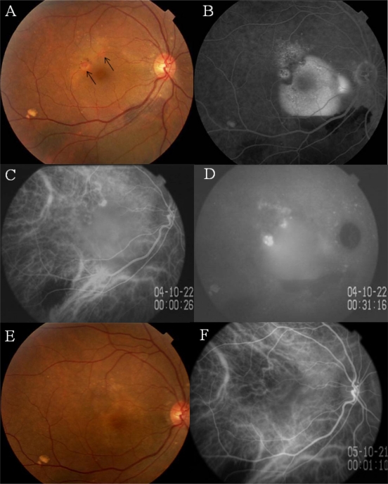
Figure 1.
Case 1. Findings in a 62-year-old monozygotic twin with polypoidal choroidal vasculopathy in her right eye.
Notes: A) Fundus photograph of right eye shows a hemorrhagic pigment epithelial detachment (PED) associated with an orange lesion beneath the retinal pigment epithelium (RPE) (arrows). B) Fluorescein angiogram shows dye pooling beneath the RPE. C) early phase indocyanine green angiogram (ICGA) showing a network of vessels and polypoidal structures. D) Late phase ICGA showing intense dye leakage from the polypoidal lesions. E) Three months after photodynamic therapy, the PED cannot be seen. F) ICGA showing closure of the polypoidal lesions.

