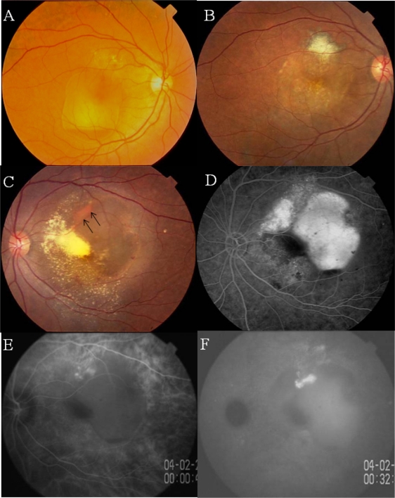
Figure 3.
Case 2. Twin sister of Case 1 with polypoidal choroidal vasculopathy in both eyes.
Notes: A, B) Fundus photographs of right eye before (A) and after (B) laser photocoagulation at the age of 57 years. C) Photograph of left fundus showing an orange lesion at the margin of a hemorrhagic PED (arrows). D) Fluorescein angiogram showing leakage of dye beneath the PED. E, F) Indocyanine green angiogram (ICGA) of right eye showing polypoidal lesions at the margin of the PED (E) with late dye leakage (F).

