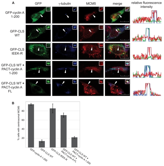
Fig. 4.
Centrosomal localization of MCM5 is dependent on cyclin A. (A) Localization of endogenous MCM5 was analyzed in CHO-K1 cells expressing either wild-type or mutant GFP-CLS, or co-expressing wild-type GFP-CLS and the indicated PACT-fused cyclin A constructs. Cells were stained with antibody to γ-tubulin (blue) and MCM5 (red). Expression and localization of GFP-tagged constructs were observed by direct fluorescence (green). Line scans measuring centrosome-associated relative fluorescence intensity are shown on the right, with the green, blue and red lines representing the GFP-, γ-tubulin- and MCM5-associated fluorescence, respectively. Arrows indicate the position of centrosomes. Insets: magnified image of the centrosomal region. Scale bars: 10 μm. (B) Graphical analysis of the centrosomal localization of MCM5. Several experiments similar to the one in A were performed with the indicated constructs. Over 100 cells were analyzed for each condition in each experiment. Error bars indicate mean ± s.d. (n=3).

