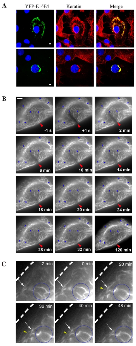
Fig. 3.
YFP-16E1^E4 associates with keratin and alters normal keratin IF network dynamics in SiHa cells. (A) Fluorescence images of SiHa cells expressing YFP-16E1^E4 (green) with a keratin immunostain (red) and DAPI stain (blue). YFP-16E1^E4 was found to associate with the filamentous keratin IF network (top panels) resulting in reorganisation of the network (bottom panels). Scale bars: 5 μm. (B) FRAP analysis of the YFP-16E1^E4-keratin network recorded over a 2 hour period, 12 hours after transfection, in SiHa cells. The first image was acquired 1 second before bleaching (−1 s); the red arrow indicates the bleached area; subsequent images were acquired after bleaching at the time points indicated. The network remained largely static over this time period (blue arrows). Fluorescence recovery occurs throughout the bleached bar, indicating association of 16E1^E4 with the polymerised keratin IF network. Scale bar: 5 μm. (C) Higher-magnification images of time-lapse frames taken at times indicated. Some peripheral filament assembly is observed (white and yellow arrows); however, on merging with the static network (blue circle) the entire network appears to contract away from the cell edge (dotted line).

