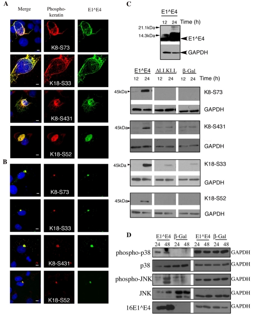
Fig. 4.
The association of 16E1^E4 with keratin results in keratin hyperphosphorylation. (A) SiHa cells infected to express 16E1^E4 were fixed and stained for 16E1^E4 (green), nuclei (blue) and phosphorylated keratin as indicated (red). In some, but not all, cells displaying filamentous 16E1^E4 staining, the associated keratin IF network is hyperphosphorylated at each of the four sites. Scale bars: 5 μm. (B) By contrast, all of the 16E1^E4 aggregate structures contain hyperphosphorylated keratin. Scale bars: 5 μm. (C) SiHa cells expressing 16E1^E4, ΔLLKLL (the 16E1^E4 keratin binding mutant) or β-Gal were harvested 12 or 24 hours after infection and analysed by western blotting using antibodies indicated on the right; GAPDH was used as a loading control. 16E1^E4 accumulated as expected over this period (top panel). Expression of 16E1^E4, but not β-Gal, results in an elevation in levels of phosphorylated keratin 8 and keratin 18. (D) SiHa cells expressing either 16E1^E4 or β-Gal were harvested 24 or 48 hours after infection and lysates analysed by western blotting using the antibodies indicated; a GAPDH loading control is also included. The levels of activated p38 MAPK and JNK are elevated in cells expressing 16E1^E4 compared with β-Gal-expressing cells.

