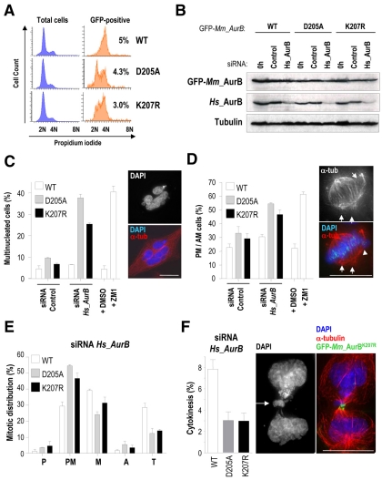
Fig. 4.
Defective mitotic progression and cytokinesis in the presence of Aurora BK207R. HeLa cells stably expressing siRNA-resistant mouse Aurora B fused to GFP were nucleofected with human AURKB siRNA oligonucleotides or a control siRNA and analyzed 36 hours after nucleofection by immunoblotting and immunofluorescence. (A) Generation and validation of HeLa stable cell lines by cytometry cell-cycle analysis. The analysis of the total cell population is shown in blue histograms, whereas the analysis of the GFP-positive population is shown in orange histograms accompanied by the percentage of these cells over the whole population. At least three different stable clones were analyzed for each GFP-Aurora-B fusion protein. Depicted histograms are from representative stable clones. (B) Western blot analysis showing efficient depletion of human endogenous Aurora B and protein levels of siRNA-resistant mouse GFP-Aurora-B proteins in HeLa stable cell lines. Tubulin was used as loading control. (C) Quantification of the percentage of interphase multinucleated cells. Stable expression of GFP-Aurora-BD205A and Aurora-BK207R induces accumulation of multinucleated cells (see picture) upon depletion of endogenous Aurora B. Around 1000 interphase cells were counted per point. (D) Quantification of the percentage of mitotic cells in prometaphase (PM) or abnormal metaphases (AM) with misaligned chromosomes (arrowheads) and multiple poles (arrows) revealing a significant arrest at these stages in cells lacking the endogenous Aurora B and stably expressing GFP-AuroraBD205A and -AuroraBK207R. About 50-100 mitotic cells were counted per point. Chemical inhibition of Aurora-B kinase activity with ZM447439 (ZM1) was included in C and D as a positive control. (E) Mitotic distribution of stable cell lines after depletion of endogenous Aurora B. Expression of GFP-Aurora-BD205A and -Aurora-BK207R provokes an arrest at prometaphase and a reduction in cells at telophase. P, prophase; PM, prometaphase; M, metaphase; A, anaphase; T, telophase. (F) GFP-Aurora-BD205A and -Aurora-BK207R stable cell lines display a reduced number of cells undergoing cytokinesis. In these mutant cells, the remaining cytokinesis figures are usually abnormal with chromosomal bridges (arrow). α-tubulin (α-tub) is in red, GFP-Aurora-B in green and DAPI (DNA) in blue. Scale bars: 10 μm.

