
FIG. 3.
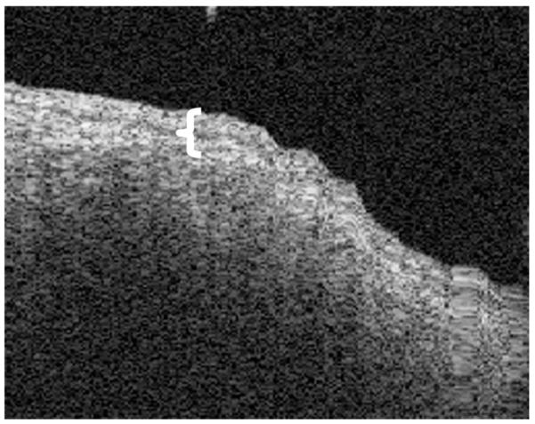
OCT image of a tympanosclerotic plaque. The image shows that the squamous epithelial layer is somewhat thicker (brackets). The calcified fibrosis prevents clear imaging of the medial layer of the TM.
Official websites use .gov
A
.gov website belongs to an official
government organization in the United States.
Secure .gov websites use HTTPS
A lock (
) or https:// means you've safely
connected to the .gov website. Share sensitive
information only on official, secure websites.
OCT image of a tympanosclerotic plaque. The image shows that the squamous epithelial layer is somewhat thicker (brackets). The calcified fibrosis prevents clear imaging of the medial layer of the TM.