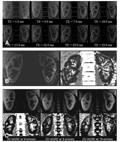
FIGURE 1.
(A) Three-dimensional multiple gradient-recalled echo images from each of the 8 echo times at the same slice position. (B) An anatomic (left) and R2* map (right) from one slice position containing a few representative regions of interest. (C) Anatomic (top row) and corresponding R2* maps (bottom row) at the same slice position for each of the 3 sequences. All images in Figure 1 were acquired at 3.0 T.

