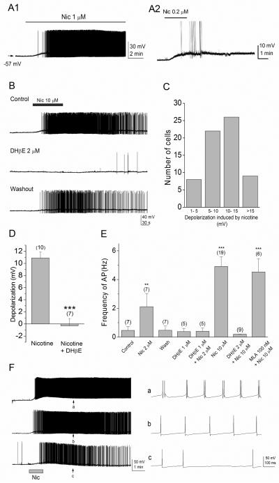
Figure 3. Nicotine depolarizes horizontally oriented interneurons in the stratum oriens/alveus and increases interneuronal spiking rate.
(A1, A2) Bath application of low concentrations of nicotine in the presence of DNQX (20 μM) and AP5 (40 μM) caused a depolarization and increased action potential firing in current-clamped interneurons. (A1) The interneurons remained depolarized during 10-min application of 1 μM nicotine. (A2) Nicotine at a low concentration found in cigarette smokers excites interneurons. (B) Bath application of 10 μM nicotine reversibly induced a depolarization of interneurons and increased the rate of action potential firing (top). These effects were blocked by 2 μM DHβE (middle). The blocking effect of DHβE was reversible after washout of the drug (bottom). (C) The magnitude of nicotine-induced depolarization varied among interneurons. (D) Summary graph showing the magnitude of depolarization observed in the presence of nicotine (10 μM) and nicotine (10 μM) + DHβE (2 μM). Note that nicotine depolarizes interneurons and the effect of nicotine was blocked by DHβE. (E) Summary graph showing the frequency of action potential observed in the absence (control) and presence of nicotine, DHβE, nicotine + DHβE, and nicotine + MLA, and after washout of nicotine (wash). Note that nicotine reversibly increased the rate of action potential firing in a dose-dependent manner, and the effect was blocked by DHβE, but not MLA. (F) Nicotine-responding interneurons exhibited different firing patterns. On the right, representative traces from three different interneurons exhibiting clustered (top), regular (middle), and irregular (bottom) firing patterns at arrows (on the left, a, b, c) are shown on an expanded time scale. **P < 0.01, ***P < 0.001.

