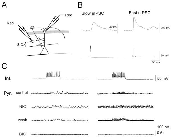
Figure 8. Nicotine-induced sIPSCs mask evoked uIPSCs originating from feedforward interneurons in the stratum radiatum in pyramidal cells.
Dual whole-cell recordings from current-clamped interneurons and voltage-clamped pyramidal cells at 0 mV were performed. (A) Schematic of recording electrode positions in the hippocampal CA1 region. (B) Slow and fast uIPSCs recorded by eliciting a spike in the interneuron by injection of a suprathreshold square current pulse. (C) On the left, a train of interneuronal spikes generated by 0.1 nA current injection evoked uIPSCs in pyramidal cells in the absence (Control) and presence of 10 μM nicotine (NIC) and after washout of nicotine (wash). All responses recorded in the pyramidal cell were blocked in the presence of bicuculline (BIC, 10 μM). On the right, five consecutive current or potential traces recorded from a pyramidal cell-interneuron pair were superimposed. Note that bath application of nicotine increased background noise and simultaneously masked evoked uIPSCs in the pyramidal cell.

