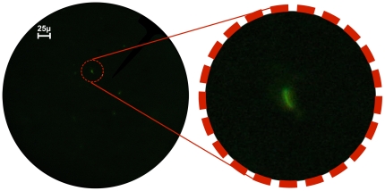
Figure 2. M. tuberculosis viewed with the Global Focus microscope.
The image on the left is a photograph of M. tuberculosis bacilli stained with auramine orange, viewed with the Global Focus microscope at 400× magnification, and captured with a Canon G9 camera (F2.8, exposure: 1 second). The image on the right is a digital magnification detail of an M. tuberculosis bacillus.

