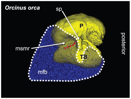
Figure 3. Lateral view of the left TPC and corresponding mandibular fat body (MFB) in Orcinus orca.
This volume has been reconstructed from CT scans of a Killer Whale (Orcinus orca) from the region around the TPC (0.3662 mm cubic voxels). As a consequence, the anterior boundary of the MFB has been artificially terminated at the anterior limit of the scanned volume. The entire head of this specimen was scanned and, as in all other odontocetes in this study, the MFB is continuous from the enlarged foramen of the mandible to its bifurcated attachment to the TPC (shown in this figure). The mandibular fat body is displayed as semi-transparent (blue), outlined in white dots, and overlies the TPC (yellow). The mallear ridge is indicated by the red dotted line. Other structures are as follows: P = periotic bone; TB = tympanic bulla; sp = sigmoid process; msmr = medial sulcus of the mallear ridge (bony funnel); mfb = mandibular fat body. The ventral branch of the MFB attaches to the tympanic bulla and the dorsal branch fits into the medial sulcus of the mallear ridge.

