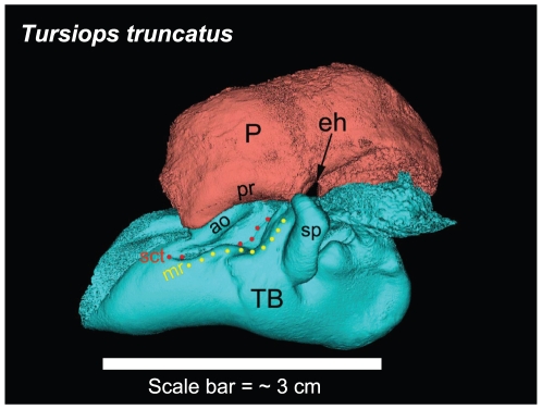
Figure 6. Lateral view of the left TPC from Tursiops truncatus reconstructed from micro-CT scans, showing major landmarks.
P = periotic bone; TB = tympanic bulla; eh = epitympanic hiatus; pr = parabullary ridge; ao = accessory ossicle; sp = sigmoid process; mr = mallear ridge (yellow dots); sct = sulcus for the chorda tympani (red dots). The scale bar represents approximately 3 cm. It is meant to give the reader an impression of the approximate size of the TPC and is not to be used to measure from point to point, considering that this is 3-D topography projected onto a 2-D plane. All TPC images of Tursiops truncatus shown in this report were reconstructed from micro-CT scans (45 micron cubic voxels).

