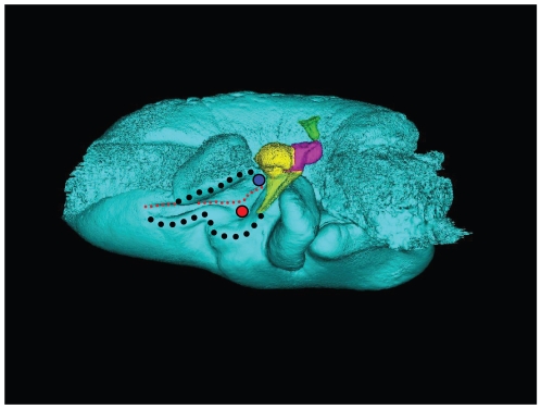
Figure 10. Dorsal view of the tympanic bone and ossicles from Tursiops truncatus.
This view is also a particularly good vantage point from which to view the structures that receive the dorsal branch of the mandibular fat body. Subtle, but potentially important, landmarks are: two very thin bony locations marked by the prominent red and blue circles. The S-shaped black dotted-line marks the mallear ridge. The shorter black dotted line marks the ridge along the accessory ossicle. The small red dotted-line marks the sulcus for the chorda tympani. The tympanic bone is colored cyan, and the ossicles are colored as follows: malleus = yellow, incus = magenta, stapes = green.

