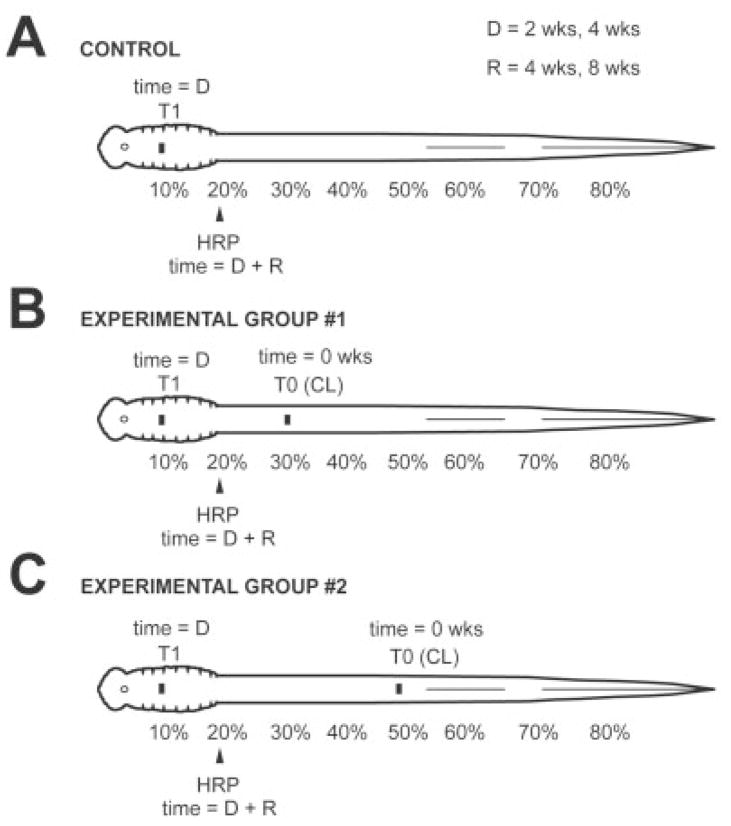
Fig. 1.
Diagrams showing experimental paradigm. A: Control group: The spinal cord was transected at 10% BL (T1), and, after recovery times of 4 or 8 weeks (R), HRP was applied to the spinal cord at 20% BL to label retrogradely descending brain neurons that regenerated their axons below the transection site. B: Experimental group 1: Animals were divided into different D-R groups, where D (2 or 4 weeks) is the lesion delay time between a conditioning lesion (T0) at 30% BL at time zero and spinal cord transection (T1) at 10% BL, and R (4 or 8 weeks) is the recovery time between spinal cord transection and application of HRP to the spinal cord at 20% BL. C: Experimental group 2: Similar to group 1, except conditioning lesions (T0) were performed at 50% BL.

