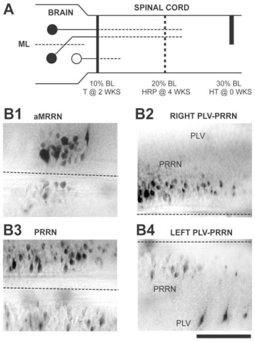
Fig. 5.
Contributions of axotomy to the conditioning lesion effect (see Table 4). A: A right hemitransection (HT) was made in the spinal cord at 30% BL, and, 2 weeks later, the spinal cord was transected (T) at 10% BL. The tracer HRP was applied to the spinal cord 4 weeks later at 20% BL. If axotomy is required for conditioning lesion effects, neurons that receive conditioning lesions (solid circles) should display enhanced axonal regeneration, whereas neurons not directly injured by the conditioning lesion (open circle) should not exhibit enhanced axonal growth. B: Photomicrographs from a brain showing asymmetrical labeling of descending brain neurons (rostral is to the left, right is upward; dashed lines indicate the midline). B1,3: There were higher numbers of labeled neurons in the right MRRN and PRRN, ipsilateral to the hemi-CL. B2,4: Larger numbers of labeled neurons were found in the right PRRN (B2) and left PLV (B4), ipsilateral and contralateral, respectively, to the hemi-CL (see solid circles in A). Scale bar = 200 μm.

