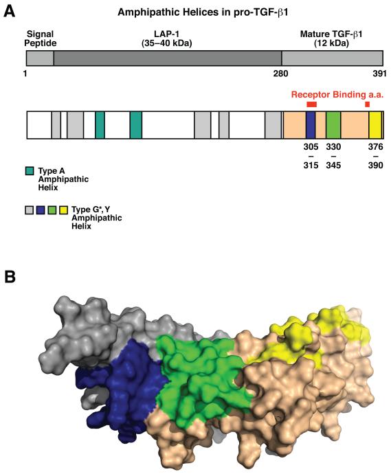
Figure 2.
TGF-β1 contains putative lipid binding domains. A, Schematic representation of lipid-binding amphipathic α-helices in pro-TGF-β1, identified by COMBO, COMMET, and CONSENSUS. Type G* or Y amphipathic helices (blue, and grey) are drawn to scale. Type G* amphipathic helices in mature TGF-β (blue, green and yellow) correspond to the same colored regions in B. B, Lipid-binding regions (blue, green and yellow) present in mature TGF-β1 form two putative lipid-binding domains. The blue region of the first chain forms a hydrophobic patch together with the green region of the second chain as shown. Grey and nude colors represent individual TGF-β1 peptides. Numbers shown correspond to amino acid numbering of pro-TGF-β.

