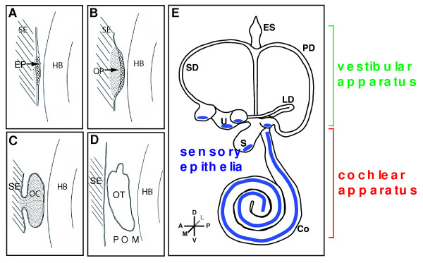
Figure 1.
Developmental milestones in mouse inner ear formation. Competence of surface ectoderm lateral to both sides of the hindbrain (HB) precedes any cell morphology changes. (A) Thickening of surface ectoderm (SE) to form the early placodes (EP) which is primarily driven by Fgf, Wnt and Pax genes. (B) Invagination of the otic placodes to form the otic pit (OP). (C) Further development and invagination of the otic pit to form the otic cup (OC) which pinches off from the surface ectoderm. (D) The separation from the overlying ectoderm gives rise to the otocyst (OT). (E) Subsequent morphogenesis to finalize the complex 3-dimensional labyrinth which is demarcated into vestibular and cochlear components. Sensory epithelia are shown in blue. Abbreviations: Co, cochlea; ES, endolymphatic sac; HB, hindbrain; LD, lateral semicircular duct; PD, posterior semicircular duct; POM, periotic mesenchyme; S, saccule; SD, superior semicircular duct; SE, surface ectoderm; U, utricle.

