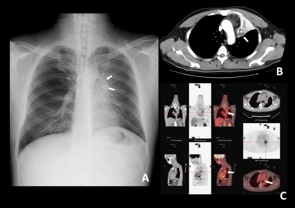
Figure 1.
(A) Chest plain film. A protruding mass shadow is seen in the left hilar region. The shadow of the left bronchus stops at the mass. Costodiaphragmatic angles are clear. There is increased density over the left lung field with elevation of the left side of the diaphragm. These findings are indicative of a hilar mass obstructing the bronchus with collapse of the left upper lobe of lung. (B) Contrast computed tomography (CT) image, distal part of the tumor. The distal bronchus is dilated and filled with secretions. The margin between the lung parenchyma and tumor is indistinct. (C) Positron emission tomography (PET) and CT, proximal part of the tumor. An endobronchial tumor with high tracer uptake and clear margins is visible.

