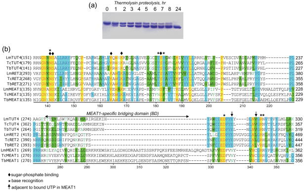
Fig. 1.
Limited proteolysis of purified recombinant MEAT1. (a) Purified MEAT1 (0.5 mg/ml) was incubated at 4° C with thermolysin (2 μg/ml) in buffer containing 50 mM Tris-HCl, pH 8.0, 100 mM KCl and 50 mM CaCl2. Aliquots were removed at indicated periods of time, separated by SDS-PAGE and stained with Coomassie blue. (b) Partial multiple sequence alignment of C-terminal domains from T. brucei TUTases with known X-ray crystal structures (RET214, TUT46 and MEAT, this study) with orthologous proteins from other Kinetoplastida species. Lm: Leishmania major; Tc: Trypanosoma cruzi; Tb: Trypanosoma brucei. TbMEAT1 amino acids are numbered; UTP-binding residues are depicted by diamonds (sugar-phosphate) and asterisks (uracil base), and arrows indicate MEAT1 positions adjacent to bound UTP.

