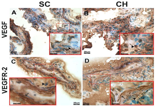
Figure 7.
IHC photomicrographs of VEGF (A, B) and VEGFR-2 (C, D) expression in choroid plexus. Cobblestone-shaped epithelial cells (arrowheads) showed similar expression of VEGF in both control (A) and hydrocephalic (B) animals. VEGFR-2 showed robust expression in the spindle-shaped endothelial cells (arrows) compared to the epithelial cells (arrowheads, C and D). When comparing control (C) and hydrocephalic (D) animals, VEGFR-2 showed similar expression. Capillary lumina are indicated by asterisks.

