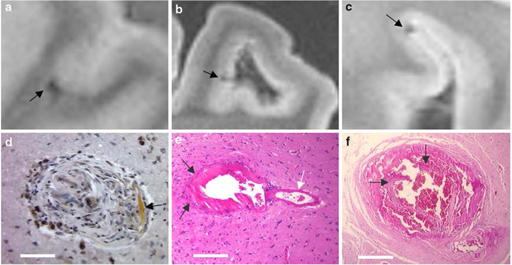
Fig. 2.
MR hypointensities without grossly visible pathology. The vast majority of hypointensities were associated with hemorrhages visible upon dissection; however, a few required more extensive investigation. Images a, b, c are MR images corresponding to the lesions shown in d, e, f, respectively. Shown in image d is a well-healed lesion consisting of focal scarring with hemosiderin deposits stained by DAB-enhanced Prussian blue stain. Hematoidin deposition is also noted in the lesion (white arrow). Image e shows an arteriolar aneurysm (arrows indicate the dome of the aneurysm; white arrow indicates the “parent” artery). Image f shows a severely dilated vessel with a dissection in the endothelium and blood in the vessel wall (arrows point the ruptured endothelial layer). Scale bars d 100 µm, e 250 µm, f 500 µm

