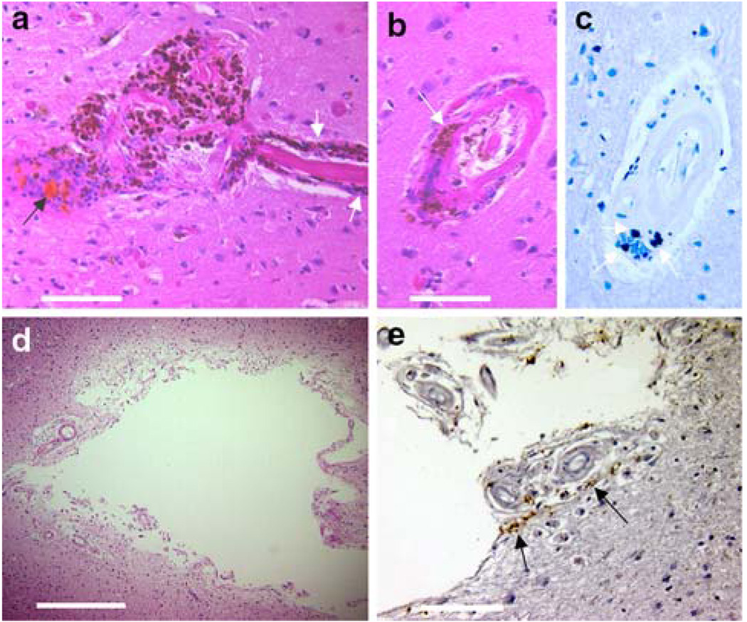
Fig. 6.
Perivascular hemosiderin deposition may contribute to subsequent ischemic changes. Image a shows a hemorrhage around a degenerated arteriole with hematoidin deposition (yellow/orange material at black arrow), significant local inflammation and hemosiderin both in the lesion and tracking in the perivascular space along the arteriole (white arrow). Image b shows another vessel, ~500 µm distant from the lesion in image a, with extensive perivascular hemosiderin (arrow). Unenhanced Prussian blue staining of the same vessel is shown in image c. Hemosiderin was present around both capillaries and arterioles at a distance of more than twice the diameter of the lesion and was accompanied by inflammatory cells. Image d shows another lesion that appeared as a hypointensity in SWI (the cavitary lesion shown in Fig. 1d–f) and on pathological examination proved to be a lacunar infarct. Image e shows DAB-enhanced Prussian blue staining of the capsule around the lesion. The vessels are also surrounded by numerous inflammatory cells and hemosiderin-laden macrophages (arrows). Scale bars a and e 200 µm, b 100 µm, d 1 mm

