
Fig. 3.
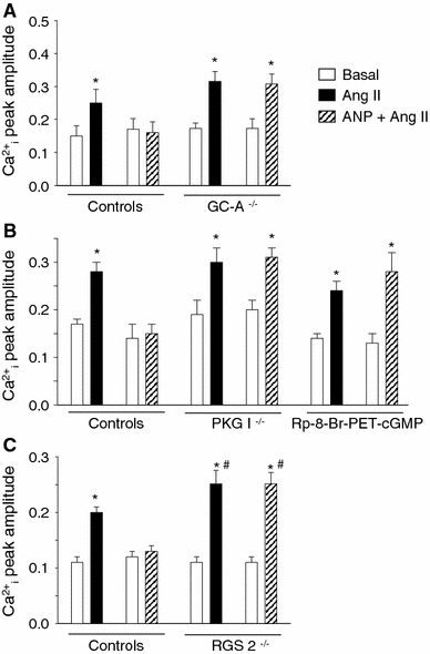
Ca2+ i transients (Indo-1 ratio405/495 nm, systolic–diastolic) in field-stimulated cardiomyocytes at baseline and during superfusion with Ang II in the presence or absence of ANP. In respective control cardiomyocytes (with unaltered protein expression levels) Ang II (10 nM) increased systolic Ca2+ i levels and the peak amplitude of Ca2+ i transients. This Ca2+ i responses to Ang II were fully prevented in the presence of ANP (100 nM, pretreatment during 10 min). This inhibitory effect of ANP on the Ca2+ i responses to Ang II was abolished a in GC-A-deficient (GC-A−/−), b in PKG I-deficient (PKG I−/−) and c in RGS2-deficient (RGS2−/−) myocytes. b The inhibitory effect of ANP on the responses to Ang II was also abolished after pharmacological blockade of PKG I with Rp-8-Br-PET-cGMP (10 μM, 30 min pretreatment). c Note that in RGS2−/− myocytes the Ca2+ i-stimulating effects of Ang II (1 nM) were significantly greater when compared with the effects of Ang II (10 nM) on control cells; n = 4–6 cells (4 mice per genotype); *P < 0.05 versus basal; # P < 0.05 versus controls