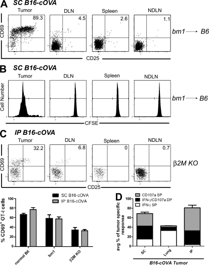
Figure 10.
Tumor cells directly presenting antigen are sufficient for the activation and effector differentiation of naive CD8 T cells. Bm1 → B6 BM chimeras (A and B) bearing established s.c., lung, or i.p. B16-cOVA tumors or β2M−/− mice bearing established s.c. or i.p. tumors (C) received Thy1-mismatched unlabeled (A and C) or CFSE-labeled (B) OT-I T cells. 24 h (A and C) or 4 d (B) after T cell transfer, tumors and lymphoid organs were harvested and lymphocytes were stained for flow cytometry. Graph in C summarizes the initial activation of OT-I cells in control mice, bm1 chimeras, and β2M−/− mice In (B) FTY720 treatment was initiated at the time of T cell transfer and continued for the duration of the experiment. All plots are gated on Thy1.1+ CD8+ OT-I lymphocytes. Numbers in (A and C) indicate the percentage of OT-I cells that are single positive for CD69 or CD25, or positive for both markers. Gates on plots in (B) indicate OT-I cells that have undergone at least one division. Axes on plots are displayed in Logicle scale. (D) Ex vivo effector analysis. OT-I cells from tumors harvested 4 d after T cell transfer were also briefly restimulated ex vivo and stained for IFN-γ and CD107a. The composition of the antitumor effector response in each tumor location is summarized in (C). Results (A, B, and D) represent n = 6 mice per tumor location and time point in 4 independent experiments, 2 with s.c. tumors and 1 each with i.p. and lung tumors. Results in C represent 3 s.c. and 3 i.p. tumor-bearing animals in 2 independent experiments.

