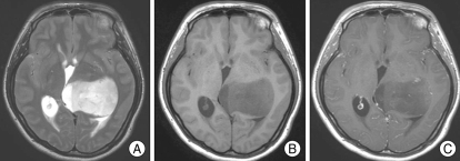
Fig. 1.
Magnetic resonance images of the tumor. A : T2-weighted image shows a well-defined high signal mass in the left thalamic pulvinar area without any involvement of the third ventricle. B : T1-weighted image shows low signal intensity of the mass. C : Gadolinium-enhanced T1-weighted image shows scattered dot-like subtle enhancement of the tumor, suggestive of the possibility of intermediate grade glioma.

