
Figure 1.
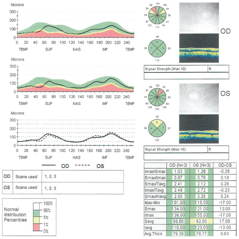
Stratus OCT printout of a peripapillary Fast retinal nerve fiber layer (RNFL) scan in a patient with bilateral glaucoma. Retinal nerve fiber layer thicknesses in the normal range are shown on green backgrounds, those that are abnormal at the 5% level are shown on yellow backgrounds, and those abnormal at the 1% level are shown on red backgrounds.