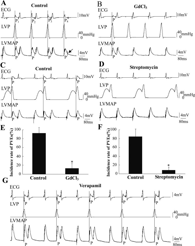
Figure 5. Effects of GdCl3 and streptomycin on pressure-induced PVEs.
A and B were recorded in the same heart before and after application of GdCl3. The LV was regularly inflated by pulses of 120 mmHg at T=180 ms from a resting pressure of 0 mmHg. In A, four numbered PVEs with four different shapes might indicate multiple PVE loci. The arrow in A indicated a pacing signal failed to pace the heart that occurred during the effective refractory period of the previous PVE. C and D were recorded before and after application of 500 μM streptomycin. The LV was regularly inflated by pulses of 120 mmHg with T=150 ms superimposed on the IPP during every other pacing cycle. The incidence of PVEs decreased after treatment with Gd3+ and streptomycin (E and F, n=14, *: P<0.01 vs. control). G shows a record when the LVP was held at 0 mmHg and stimulated with 60-mmHg pulse for T=180 ms after application of 1 μM verapamil. The PVEs caused by pressure pulses could not be blocked by verapamil in five hearts.

