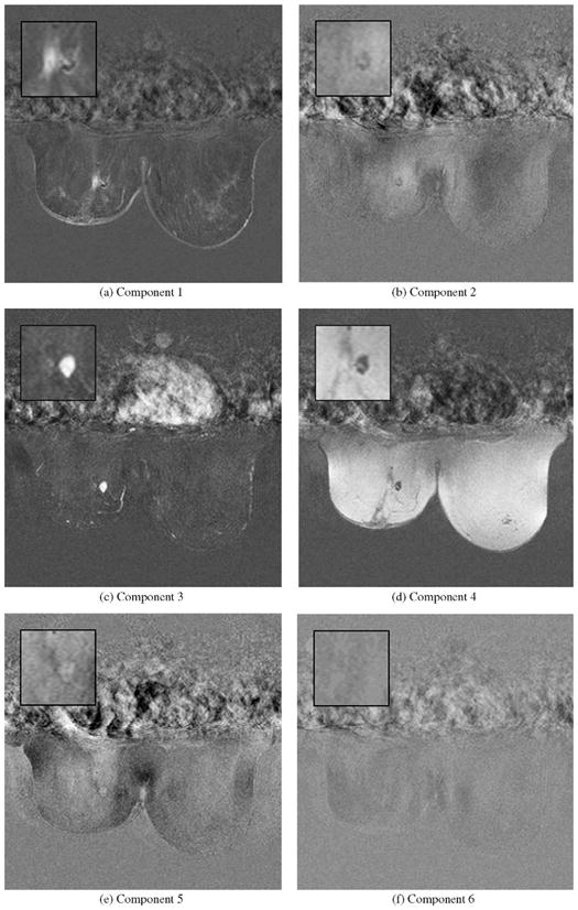
Fig. 5.

Slice based representation of six score images for case XII based on Icasso (Tanh). In the first image the location and extent of one lesion compartment could be identified which exhibits a continuously increasing signal whereas blood vessels with a similar characteristic become also visible. In the third image another compartment of the lesion stands out which shows a typical uptake-washout pattern which is highly indicative for malignant lesions. (a) Component 1. (b) Component 2. (c) Component 3. (d) Component 4. (e) Component 5. (f) Component 6.
