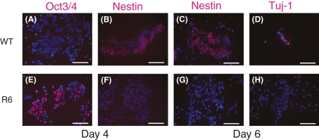
Figure 4.
Comparison of marker protein expression under a neural differentiation condition. The control (WT, upper panels) and Hes1-sustained cells (R6, lower panels) cultured in N2B27 medium were analyzed on days 4 and 6 by immunocytochemistry using anti-Oct3/4, anti-Nestin and anti-Tuj-1 antibodies (red) with DAPI staining (blue). Scale bars, 100 μm.

