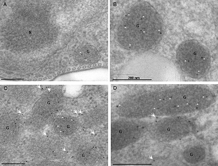
Fig. 7.

Localization and distribution of GAPDH by immuno-gold labelling. Thin-layer sections were labelled with anti-GAPDH and stained with protein A gold particles and examined by TEM. WT (A), RNAi non-induced (B) and induced (C–D) at 72 h. Abbreviations used: nucleus (N); glycosome (G) and white asterisks alongside black dots represent 10 nm gold particles present within glycosomes, while white arrows depict particles outside glycosomes. Scale bar represents 200 nm.
