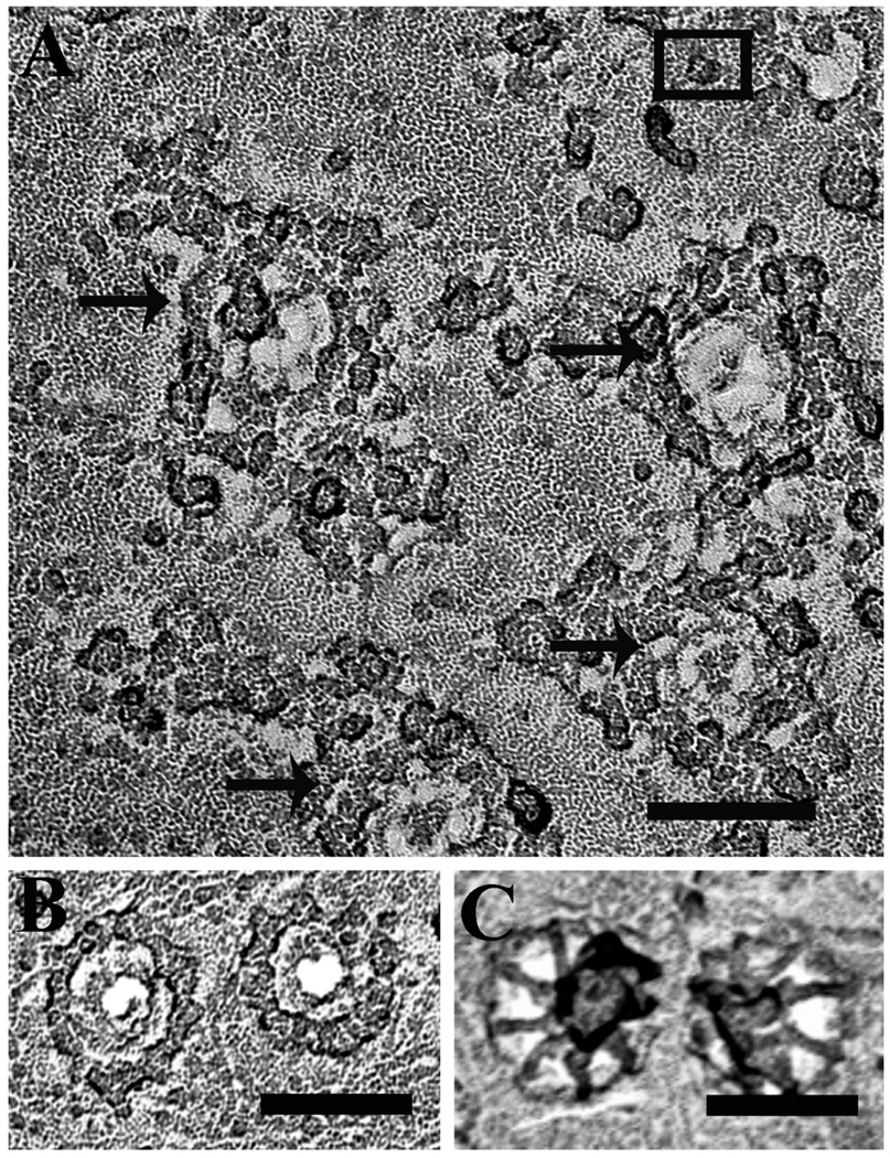
Figure 1. Identification of nuclear envelope surfaces in isolated nuclei by freeze-drying and rotary shadowing.
A. Cytoplasmic surface of a nucleus isolated from wild-type Sf9 cells. The nuclear pore complexes (NPC), arrows) are identifiable as annuli with a diameter of 100–110 nm, decorated by a ring of prominent particles. The structure is typical of the cytoplasmic side of the envelope [36] and intact nuclei always showed only this side The black square encloses a particle identified as a presumptive InsP3R based on evidence shown later. Scale bar = 100 nm. B and C. Details of freeze-dried rotary-shadowed NPCs in the cytoplasmic (B) and interior face (C) of nuclear envelope fragments from Xenopus oocytes Note that in B the NPC is dominated by a particulate ring, while in C the dominant structure is a fibrillar basket.

