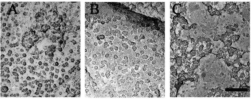
Figure 3. Detection of InsP3R in rotary shadowed images of isolated nuclei.
Nuclei isolated from Sf9 cells expressing either rat type 1 IP3R (A), or type 3 IP3R (B), or Wolframin, a non-related ER membrane protein, (C) were freeze-dried and rotary shadowed. Nuclear membranes from type 1 and type 3 InsP3R-expressing cells showed large membrane patches occupied by a high density of small structures ~15 nm on the side similar to the rarer particles observed in the uninfected cell nuclear membranes. Nuclear pores were absent from these patches but visible elsewhere in the same nuclei. The small particles were not visible in cells transfected with cDNA for Wolframin. Scale bar, 100 nm.

