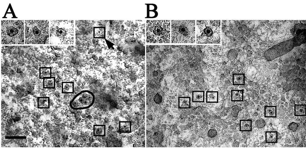
Figure 5. Shadowed images of nuclear envelopes from Sf9 cells expressing rat type 3 InsP3R.
The nuclei were treated either with gold-labeled heparin (A) or with anti-type 3 InsP3R followed by gold-labeled secondary antibody (B) followed by freeze-drying and rotary shadowing (see Methods). In both cases, the 5 nm gold particles are clearly associated with the InsP3R-like structures, with the exception of 1 out of 11 gold particles in (A) (arrow). High magnification (insets) shows the gold particles mainly located in the central region of the structures. Note that the InsP3R-like structures labeled by the gold particles have similar sizes but variable shapes and overall resemble the presumptive InsP3R particles described above. The images are not as sharp as those in the previous figures because the technique used leaves some cellular debris associated with the shadowed replicas.

