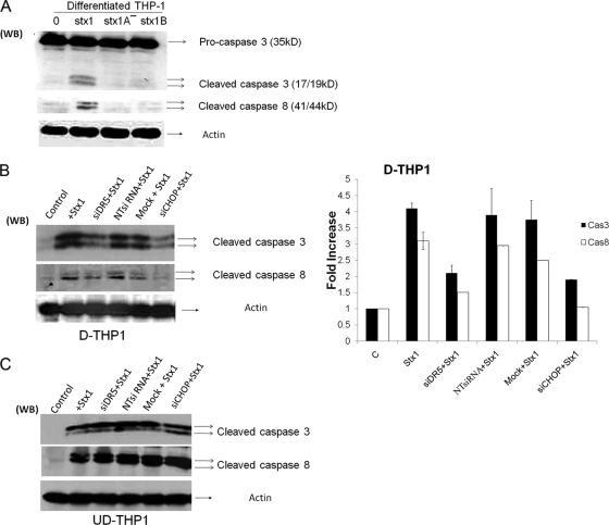
FIG. 6.
Caspase activation following Stx1 treatment of DR5 or CHOP siRNA-transfected macrophage-like and monocytic THP-1 cells. (A) Differentiated THP-1 cells were treated with Stx1, Stx1A−, or Stx1 B subunits for 8 h. Cells were lysed, followed by Western blotting with anti-caspase-3 and -8 antibodies. Membranes were then stripped and reprobed with antiactin antibody for an equal-protein-loading control. (B) Differentiated THP-1 cells (D-THP1) were transfected with siRNAs specific for DR5 (siDR5), CHOP (siCHOP), or nontargeting siRNA (NTsiRNA) or were mock transfected (Mock) with reagent only for 72 h. After washing, cells were incubated with or without Stx1 for 8 h. Cell lysates were analyzed for caspase-3 and -8 cleavage as described above. A representative Western blot is shown in the left panel; the bar graph (right panel) depicts results from at least three independent experiments with caspase activation expressed as fold increase compared to control (untreated) cells. (C) Undifferentiated THP-1 cells (UD-THP1) were transfected with siRNAs and treated with Stx1 as outlined above. A representative Western blot showing caspase-3 and -8 cleavage is shown.

