FIG. 5.
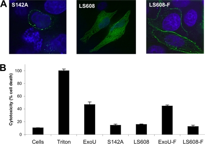
Localization and cytotoxicity of farnesylated ExoU proteins. To test the effects of farnesylation on ExoU function, GFP-tagged ExoU proteins were expressed in HeLa cells by transient transfection. At 20 h posttransfection, cells were analyzed for ExoU localization and for cytotoxicity. (A) To assess localization, cells were fixed, treated with Hoechst stain to label nuclei (blue), and visualized by fluorescence microscopy. Comparison of unfarnesylated protein (GFP-ExoU-S142A and GFP-ExoU-LS608) and farnesylated protein (GFP-ExoU-LS608-F) demonstrated that farnesylation resulted in localization of protein to the plasma membrane. Representative images are shown. (B) The cytotoxicity of ExoU variants was quantified by measuring LDH release. Triton was used to cause 100% cell lysis. Experiments were performed in triplicate; data represent means ± standard deviations. Cells, no transfection; LS608-F, GFP-ExoU-LS608 containing a C-terminal farnesylation sequence; ExoU, GFP-ExoU; S142A, GFP-ExoU-S142A, etc.

