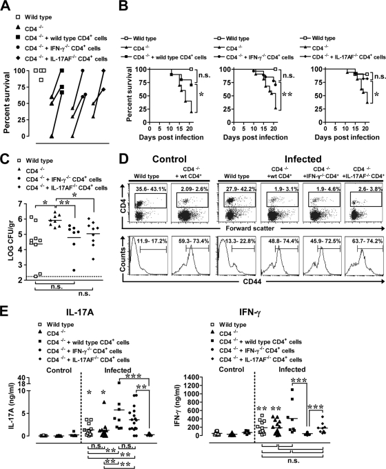
FIG. 8.
Adoptively transferred wild-type, IFN-γ−/− or IL-17AF−/− CD4+ T cells improve survival of neonatal CD4−/− mice following infection with Y. enterocolitica, and MLN cells from these mice produce inflammatory cytokines upon in vitro stimulation with Y. enterocolitica antigens. Lymph node CD4+ T cells (6 × 10⁶) from adult C57BL/6, IFN-γ−/−, or IL-17AF−/− mice were injected intravenously into 1-day-old CD4−/− mice. (A) Seven-day-old wild-type C57BL/6, CD4−/−, CD4−/− mice injected with wild type CD4+ cells, CD4−/− mice injected with IFN-γ−/− CD4+ cells, and CD4−/− mice injected with IL-17AF−/− CD4+ cells were infected orogastrically with 1 × 10⁷ CFU of Y. enterocolitica. The percent survival values from two to three individual experiments are connected with a line. (B) The data from the experiments in panel A were pooled to generate survival curves and analyzed by the Mantel-Haenszel log rank test. n.s., not significant; *, P ≤ 0.03; **, P = 0.0025 (for CD4−/− versus CD4−/− mice injected with CD4+ cells). (C) The small intestines were collected at 25 days p.i. from infected wild-type, CD4−/−, CD4−/− mice injected with IFN-γ−/− CD4+ cells, or CD4−/− mice injected with IL-17AF−/− CD4+ cells, and tissues analyzed for Yersinia titers. The dashed line is at the limit of detection. The data from two experiments were pooled (n = 7 to 9 mice per group) and analyzed by the Mann-Whitney test. n.s., not significant; *, P = 0.01; **, P = 0.0012. (D) MLN from mice that survived the infection at 21 to 25 days p.i. were collected, and cell suspensions were stained with anti-CD4 and anti-CD44 antibodies. Representative flow cytometry profiles show the percentages of CD4+ (top) and CD44+ cells within CD4+ cells (bottom), with the range of cells from three to four experiments (n = 5 to 17 mice). (E) MLN cells from individual mice described in panel D were cultured for 72 h with 10 μg/well of HKY. MLN cells from uninfected C57BL/6 (n = 8), CD4−/− (n = 25), and CD4−/− mice injected with wild-type CD4+ cells (n = 5) were stimulated along with cells from infected CD4−/− (n = 15), CD4−/− mice injected with wild-type CD4+ cells (n = 8), CD4−/− mice injected with IFN-γ−/− CD4+ cells (n = 17), and CD4−/− mice injected with IL-17AF−/− CD4+ cells (n = 9). Supernatants were tested for IFN-γ and IL-17A by ELISA. Differences were analyzed by the Mann-Whitney test. n.s., not significant; For IL-17A, significance is indicated as follows: *, P ≤ 0.013 between infected CD4−/− plus IL-17AF−/− CD4+ cells versus infected wild-type or infected CD4−/− mice; **, P ≤ 0.0049, ***P ≤ 0.0004 (between selected groups). For IFN-γ, significance is indicated as follows: **, P ≤ 0.0025 between infected CD4 −/− plus IFN-γ−/− CD4+ cells versus infected wild-type or infected CD4−/− mice; ***, P ≤ 0.0004 (betwen selected groups).

