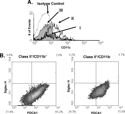
FIG. 3.
Dendritic cell staining in Toxoplasma-infected eyes. Mice were intravitreally injected with 104 parasites. Six days later, eyes were harvested and analyzed using flow cytometry. (A) Histogram of CD11c expression on the three populations of MHC class II-expressing cells from Fig. 2A. (B) FACS plots of PDCA1 and Siglec-H staining of class II+ CD11b+ and class II+ CD11b− cells.

