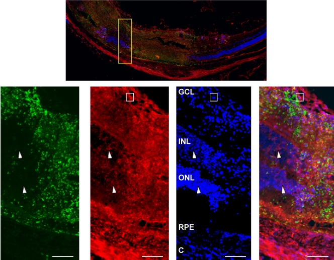
FIG. 4.
MHC class II is expressed on resident retinal cells in Toxoplasma-infected retinas. Mice were intravitreally injected with 104 parasites. Six days later, eyes were harvested, fixed, and processed for immunocytochemistry. Sections were stained to detect either DAPI (blue), MHC class II (red), or SAG1 (green). (Top) Low-magnification (×40) image of the posterior retina from an infected eye. (Bottom) High-magnification (×400) images of the area highlighted in the yellow box in the top image. Note the SAG1 staining and the intense class II staining in necrotic regions of the retina. Arrowheads point to class II+ nuclear cells in inner and outer nuclear layers. The boxes highlight a crescent-shaped, class IIhi monocyte. Bars, 100 μm. Abbreviations: C, choroid; RPE, retinal pigmented epithelium; ONL, outer nuclear layer; INL, inner nuclear layer; GCL, ganglion cell layer.

