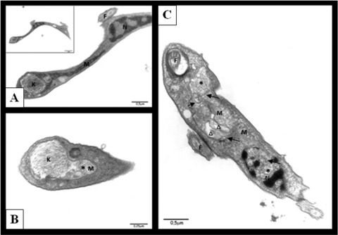
FIG. 8.
Ultrastructural changes in 7a-treated trypomastigotes. (A) Control parasite: normal mitochondrion (M), nucleus (N), kinetoplast (K), and flagellum (F); (B and C) treated parasites, showing mitochondrion swelling (asterisk), formation of abnormal membrane structures inside the organelle (black arrows), and atypical vacuoles (triangle).

