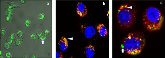
FIG. 2.
Confocal microscopy. (a) Uptake of green PNFs in J774A.1 cells. (b) Nanostructures are shown by yellow-to-orange spots formed by green nanoparticles/dyes and red endosomes/lysosomes, showing that a majority of the Alexa Fluor appears to reside in endosomes (arrowhead). (c) PNFs are distributed throughout the cells. Colocalization of PNFs with endosomes/lysosomes after incubation for 2 h (arrowhead) and subcellular localization of PNFs (arrow).

