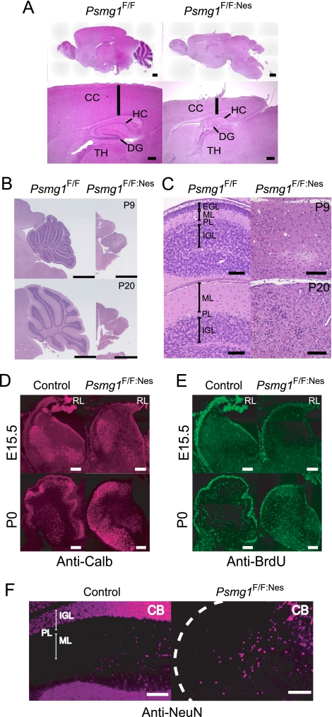
FIG. 3.
Disorganized brain structure in Psmg1F/F:Nes mice. (A) Sagittal sections of Psmg1F/F and Psmg1F/F:Nes forebrains at P21 were stained with hematoxylin-eosin. The sections were observed at low (upper panel; scale bar, 600 μm) and high (lower panel; scale bar, 300 μm) magnification. CC, cerebral cortex; HC, hippocampus; TH, thalamus; DG, dentate gyrus. (B) Histological analysis of Psmg1F/F and Psmg1F/F:Nes cerebella at P9 and P20. A section of each brain was stained with hematoxylin-eosin. Scale bar, 1 mm. (C) High-magnification images of images from panel B. Scale bar, 100 μm. EGL, external granule layer; ML, molecular layer; PL, Purkinje cell layer; IGL, internal granule layer. (D) Immunofluorescent staining of Purkinje cells in the cerebellum at E15.5 and P0 by anticalbindin antibody. (E) BrdU staining following BrdU injection into E15.5 pregnant mice and P0 mice. (F) Mature neurons in the cerebellum at P21 were visualized by immunofluorescent staining against NeuN. Abbreviations are the same as those for panel B.

