FIG. 5.
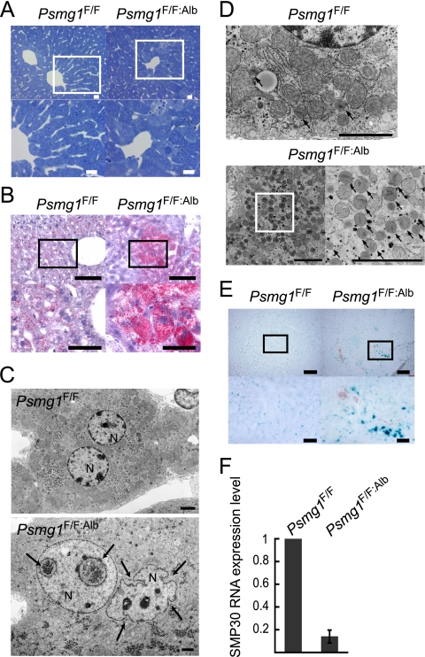
Premature senescence-like phenotypes in Psmg1F/F:Alb liver. (A) Toluidine blue staining of Psmg1F/F and Psmg1F/F:Alb livers at 6 months of age. The lower panels are higher-magnification images of the regions outlined by white rectangles in the upper panels. Scale bars, 20 μm. (B) Cryosections of 1-year-old livers were stained with oil red O. The lower panels are higher-magnification images of the regions outlined by the rectangles in the upper panels. Scale bars, 100 μm (upper panels) and 50 μm (lower panels). (C) Electron-micrographic examination of hepatocyte nuclei in Psmg1F/F and Psmg1F/F:Alb livers at 6 months of age. The invagination of the nuclear envelope is indicated by arrows. N, nucleus. Scale bar, 2 μm. (D) Electron micrographs of hepatocyte peroxisomes in Psmg1F/F:Alb livers at 6 months of age. The lower right panel is a higher-magnification image of the region outlined by the rectangle in the lower left panel. Scale bar, 2 μm. (E) Detection of senescence-associated β-galactosidase activity on cryosections of Psmg1F/F and Psmg1F/F:Alb livers at 1 year of age. The lower panels are higher-magnification images of the regions outlined by the rectangles in the upper panels. Scale bar, 200 μm (upper panels) and 50 μm (lower panels). (F) Expression of senescence marker protein 30 (SMP-30) mRNA. The relative amount of SMP-30 mRNA in Psmg1F/F:Alb liver at 3 months of age was measured by real-time PCR analysis. The data represent means ± standard deviations (SD) from three independent experiments.

