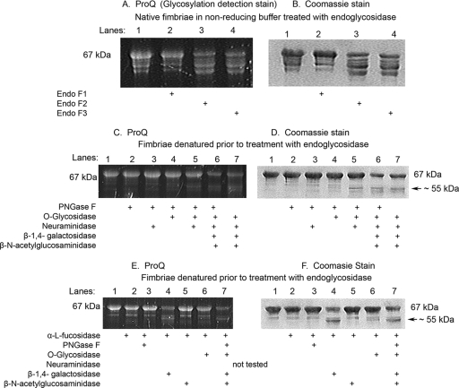
FIG. 3.
Enzymatic deglycosylation of the minor fimbria observed in the presence of endoglycosidase F2, endoglycosidase F3, and β(1-4)-galactosidase. Enzymatic deglycosylation treatment on purified minor fimbria (Mfa1), as verified by lack of shift or the loss of ProQ (glycosylation detection) signal, is shown. (A, C, E) ProQ gels; (B, D, and F) the same gel after Coomassie blue staining. (A and B) Nonreduced native fimbria treated with endoglycosidase. All lanes were loaded with 5 μg of Mfa1 and digested with the indicated endoglycosidase(s). (C and D) Fimbriae denatured prior to treatment with endoglycosidase. All lanes were loaded with 7 μg of Mfa1 and digested with the indicated endoglycosidase(s). (E and F) Minor fimbria pretreated with α-l-fucosidase, then denatured and treated with endoglycosidase. All lanes were loaded with 7 μg Mfa1 and digested with the indicated endoglycosidase(s).

