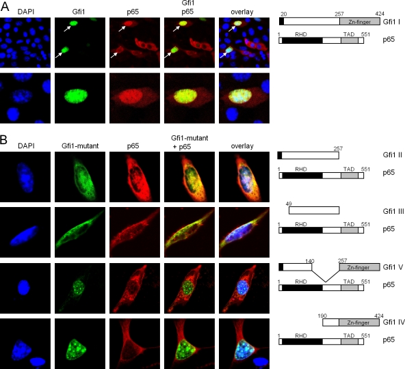
FIG. 7.
Gfi1 colocalizes with p65 in the nucleus. (A) NIH 3T3 fibroblasts were transfected with constructs directing the expression of full-length Gfi1 as a fusion protein with GFP or a full-length Myc-tagged p65 protein. Nuclei were visualized by DAPI (4′,6-diamidino-2-phenylindole) staining, and p65 was visualized by staining with anti-Myc antibodies and rhodamine-labeled secondary antibodies. Cells were analyzed with a laser scanning microscope (LSM). The merged pictures demonstrate colocalization of Gfi1 (green) and p65 (red) in cells that coexpress both proteins (white arrows). (B) Cotransfection of the indicated expression constructs, as described for panel A, that allow the production of either full-length Myc-tagged p65 or the Flag-tagged Gfi1 II, III, V, and IV mutants. Gfi1 mutants II and III show a partial colocalization with p65 (first and second rows), but Gfi1 mutants IV and V showed no colocalization (third and fourth rows).

