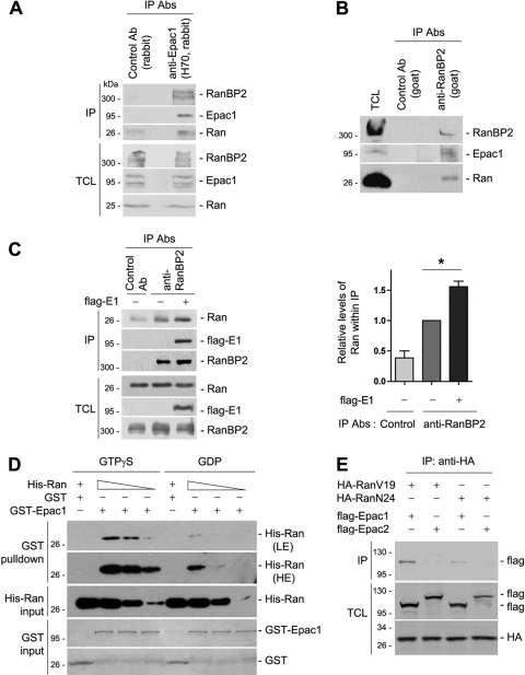
FIG. 1.
Association of Epac1 with Ran and RanBP2. (A) Coimmunoprecipitation (IP) of endogenous Epac1 with Ran and RanBP2 in HEK293 cells. Cell lysates were subjected to IP using anti-Epac1 (H70) and unrelated rabbit IgG as a control. Western blotting was performed using anti-RanBP2 (Novus), anti-Epac1 (H70), and anti-Ran antibodies (Abs). TCL, total-cell lysates. (B) Coimmunoprecipitation of endogenous RanBP2 with Epac1 and Ran in HEK293 cells. Cell lysates were subjected to IP using an anti-RanBP2 antibody (goat) and unrelated goat IgG as a control. The presence of RanBP2, Ran, and Epac1 within the IP was analyzed by Western blotting using anti-RanBP2 (Novus), anti-Epac1 (A5), and anti-Ran. (C) Effect of Epac1 overexpression on the association of RanBP2 and Ran. IP with anti-RanBP2 was performed as for panel B in the presence (+) or absence (−) of Flag-Epac1 (E1), and the presence of Ran, RanBP2, and Flag-Epac1 within the IP was determined by Western blotting. TCL were also determined by Western blotting. The left panel shows one representative result, and the right panel shows the quantification of the results of five independent experiments normalized to the level of Ran seen in the RanBP2 IP in the absence of transfected Epac1 (means ± standard errors of the means; *, P < 0.05). (D) GTP-dependent interaction between Epac1 and Ran in vitro. Increasing amounts of His-Ran loaded with GTPγS or GDP were incubated with GST or GST-Epac1 and were detected by Western blotting using anti-Ran. LE and HE, low and high exposures. GST and GST-Epac1 levels were shown with Coomassie blue. (E) Association of Ran with Epac1 but not Epac2. HA-RanV19 or HA-RanN24 was coexpressed with Flag-tagged Epac1 or Epac2 in HEK293 cells. IP was performed using anti-HA, and Western blotting was performed using anti-Flag and anti-HA. Data shown are representative of at least three independent experiments.

