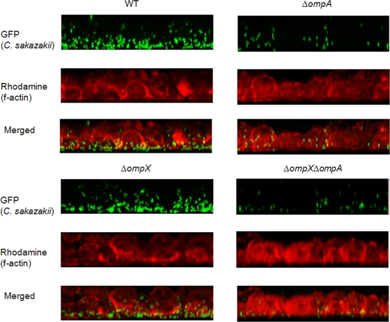
FIG. 7.
Confocal fluorescence microscopy of Caco-2 cells that were EGTA treated and infected with C. sakazakii strains containing pWM1007 for 30 min. The f-actin molecules were stained with rhodamine-labeled phalloidin. Different series of images from 1-μm x-y-z sections were obtained, analyzed, and stacked using the recommended software (LSM-FCS).

