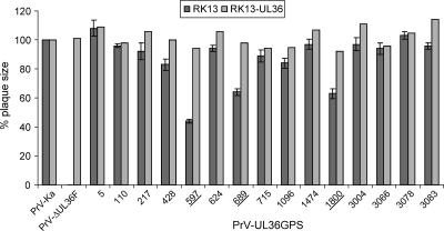
FIG. 3.
Determination of plaque size. For analysis of plaque diameters, nontransgenic and pUL36-expressing RK13 cells were infected under plaque assay conditions. Cells were fixed 2 days p.i., and plaque diameters were measured microscopically. For each virus, 50 plaques were measured in three independent experiments on RK13 cells and once on complementing cells. Values were calculated compared to wild-type PrV-Ka, which was set at 100%. Standard deviations are indicated where appropriate. UL36GPS insertion mutants which show a significant decrease in plaque diameter are underlined.

