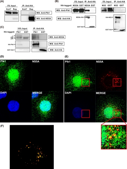
FIG. 4.
Plk1 associates and partially colocalizes with NS5A. (A) Coimmunoprecipitation of replicon NS5A and endogenous Plk1. Lysates from Huh-7 and HCVrep-HA replicon cells were collected and immunoprecipitated with anti-HA agarose, and then the eluate was subjected to Western blotting with anti-Plk1 and anti-NS5A antibodies. (B) 293T cells were cotransfected with pUI-Plk1 and NS5A-HA-, HA-NS3/4A-, or GST-HA-expressing vectors. At 48 h posttransfection, immunoprecipitation and Western blotting were performed as above. Anti-HA was used for NS proteins and GST detection. (C) Western blot analysis of HA-Plk1 immunoprecipitates. 293T cells were cotransfected with pCI-HA-Plk1/pCI-GST-HA and pUI-NS3-5B. At 48 h posttransfection, immunoprecipitation and Western blotting were performed similar to the experiments described in panels A and B. IP, immunoprecipitation; WB, Western blotting. (D and E) Immunohistochemical staining of NS5A-HA and Plk1 in Huh-7 cells (D) and HCVrep-HA replicon (E). Cells were fixed and stained at 2 days after subculture. Green, anti-Plk1; red, anti-HA (NS5A); blue, DAPI (nucleus). (F) The image analysis of colocalization coefficient using the Zen 2009 program provided with the LSM710 confocal microscope (Zeiss). Red, NS5A signals not colocalizing with Plk1; yellow, NS5A signals colocalizing with Plk1.

