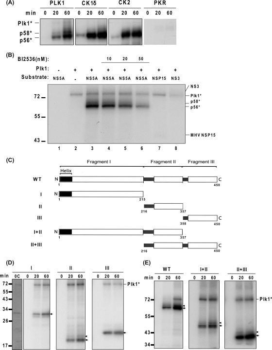
FIG. 5.
NS5A is hyperphosphorylated in vitro by Plk1. (A) In vitro kinase assay. His-tagged NS5A proteins were expressed in E. coli and purified with Ni-affinity gel and dialyzed. These purified NS5A proteins were then incubated with the kinases as indicated in the presence of [γ-32P]ATP. Reaction time ranged from 0 min to 60 min. (B) The specificity of Plk1 kinase. BI2536 treatment and negative-control proteins, MHV nsp15 and HCV NS3, were indicated. The reaction mixture was incubated for 60 min. (C) Schematic presentation of the NS5A derivatives used in this study. The diagram of the full-length NS5A structure is modified from Tellinghuisen et al. (51). The N-terminal and C-terminal amino acids of each fragment are labeled below. (D and E) Incubation of His-tagged truncated NS5A proteins with Plk1 in vitro. The basal phosphorylated forms corresponding to the size of unphosphorylated fragments are indicated with arrows; the hyperphosphorylated forms are denoted by arrowheads. Reaction time ranged from 0 min to 60 min as indicated. 0C, Coomassie blue staining of the proteins at 0 min of the reaction. Plk1*, autophosphorylated Plk1. Molecular sizes are indicated on the left (in kDa).

